

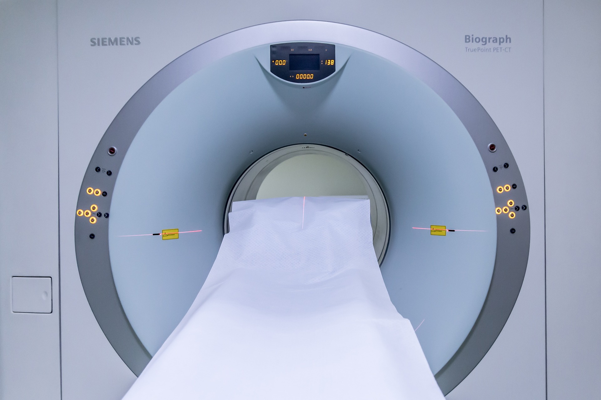
A standard MRI captures sharp, static pictures of your anatomy, while an fMRI tracks metabolic activity in your brain over time. Understanding the difference between FMRI and MRI helps clarify why doctors pick one scan for physical injuries and the other to map active brain circuits. Both use strong magnetic fields, yet they solve fundamentally different medical problems.
Table of Contents
| Feature | Standard Structural MRI | Functional MRI (fMRI) |
|---|---|---|
| Primary Goal | Maps static anatomical structure | Tracks dynamic metabolic function |
| Output | High-resolution 2D and 3D anatomical images | Color-coded brain activity maps over time |
| Signal Source | Hydrogen proton density and tissue relaxation times | Blood-Oxygen-Level-Dependent (BOLD) contrast |
| Key Resolution | High spatial resolution (sub-millimeter) | Higher temporal resolution (seconds per volume) |
| Patient Role | Must remain completely still and passive | Performs active mental tasks or rests during scanning |
What Is the Core Difference Between FMRI and MRI?
The fundamental difference between FMRI and MRI comes down to anatomy versus activity. A standard magnetic resonance imaging scan acts like a high-definition camera for your internal tissues. It reveals physical shapes, tissue boundaries, and lesions clearly. In contrast, functional magnetic resonance imaging works more like a video camera tracking blood flow changes across your brain.
Doctors order standard scans to inspect physical structures like torn ligaments, spinal discs, or brain tumors. Meanwhile, researchers and neurosurgeons use functional imaging to see which specific brain regions activate when you speak, move, or think. This essential difference between FMRI and MRI determines how clinical teams approach neurological care.
Structural MRI: Capturing Static Anatomy
Standard MRI produces detailed anatomical pictures of organs, soft tissues, and bones without using ionizing radiation. The scan provides exceptional spatial resolution, showing fine tissue boundaries down to fractions of a millimeter. Consequently, radiologists rely on these clear images to spot physical damage, inflammation, strokes, and structural anomalies.
Because the image represents a static snapshot, it shows what the brain looks like rather than what it is currently doing. A healthy brain scan and a depressed patient’s brain scan might look identical on a regular MRI if no physical tissue damage exists. This distinction highlights a major difference between FMRI and MRI in modern diagnostics.
Functional MRI (fMRI): Tracking Metabolic Activity
Functional MRI detects dynamic physiological changes in brain tissue over time, marking a central difference between FMRI and MRI. When your brain works on a task, active neurons demand more energy and oxygen. The scanner detects these localized vascular shifts across seconds of activity.
Instead of merely producing a black-and-white anatomical picture, functional scans generate statistical maps of neural activation. Technicians overlay these vibrant color maps directly onto structural images. As a result, clinicians can see the exact physical location of active cognitive processes.
How the Underlying Technology Differs
Both machines use powerful magnetic fields, typically measured at 1.5 Tesla (1.5T) or 3 Tesla (3T). Yet the mathematical sequences and physical signals they measure differ significantly. Understanding the technical difference between FMRI and MRI requires looking at how hydrogen atoms and blood molecules react inside a magnetic field.
How Standard MRI Maps Tissue Contrast
The human body consists mostly of water and fat, which contain abundant hydrogen protons. Inside an MRI scanner, the powerful magnetic field aligns the spin axes of these protons. The machine then pulses radiofrequency waves to knock these protons out of alignment.
When the radio pulse stops, protons relax back to their baseline alignment and emit faint radio signals. Different tissues, such as gray matter, white matter, cerebrospinal fluid, and bone, relax at distinct speeds. The machine calculates these relaxation times (known as T1 and T2 relaxation) to construct detailed anatomical slices with sharp tissue boundaries.
How fMRI Uses the BOLD Signal to Detect Neural Activity
Functional imaging relies on a biological phenomenon known as the hemodynamic response. When neurons fire, local blood vessels dilate to supply those active brain regions with fresh, oxygenated blood. This surge creates the Blood-Oxygen-Level-Dependent (BOLD) contrast signal.
The physics behind this process depends on hemoglobin properties, as detailed in comprehensive guides from the National Institute of Biomedical Imaging and Bioengineering. Oxygenated hemoglobin (oxyhemoglobin) is diamagnetic and does not disrupt magnetic fields. In contrast, deoxygenated hemoglobin (deoxyhemoglobin) is paramagnetic and creates tiny magnetic distortions that attenuate the MRI signal.
When an active brain area receives a rush of oxygen-rich blood, local deoxyhemoglobin levels drop quickly. This shift enhances the local MR signal intensity. Tracking these tiny signal fluctuations over time is the key technical difference between FMRI and MRI.
Spatial vs. Temporal Resolution Trade-Offs
Every neuroimaging method faces a direct trade-off between spatial and temporal resolution. This balance represents another practical difference between FMRI and MRI scans.
- Spatial resolution: Standard structural scans excel here. They capture details below one millimeter, letting physicians see minute tissue lesions clearly.
- Temporal resolution: Functional scans sacrifice spatial sharpness to capture rapid brain changes every one to two seconds.
Because functional scans must capture dynamic physiological changes quickly, each individual image frame has lower spatial detail. Radiologists overcome this limitation by superimposing low-resolution functional activation maps over high-resolution structural scans.
When Is an fMRI Used Instead of a Regular MRI?
Doctors do not simply substitute one scan for the other. Instead, the clinical question dictates the choice. Knowing the practical difference between FMRI and MRI helps explain why standard scans dominate routine hospital orders while functional scans remain specialized.
Common Clinical Uses for Structural MRI
Hospitals run millions of standard MRI scans every year because they assess physical tissue integrity across the entire body. Common clinical uses include:
- Identifying acute ischemic strokes, cerebral hemorrhages, and vascular aneurysms.
- Diagnosing structural brain tumors and tracking physical tumor growth over time.
- Detecting multiple sclerosis plaques and white matter lesions.
- Evaluating spinal disc herniations, joint cartilage tears, and ligament damage.
In each scenario, the physician needs to see physical damage or structural abnormalities. A standard structural scan answers these anatomical questions quickly and reliably.
Pre-Surgical Brain Mapping and Cognitive Research for fMRI
Clinicians turn to functional imaging when they need to preserve vital brain functions before surgery. If a neurosurgeon must remove a tumor near speech or motor centers, an fMRI maps those critical eloquent areas beforehand. The surgical team can then plan a safe path that avoids permanent neurological deficits.
Beyond surgical suites, cognitive neuroscientists use functional scans to study human cognition. Researchers track active brain networks during language processing, sensory perception, emotional regulation, and memory retrieval. Understanding this research utility clarifies another major difference between FMRI and MRI in modern neuroscience.
Task-Based vs. Resting-State Functional Imaging
When comparing scanning protocols, the difference between FMRI and MRI becomes evident in how testing paradigms are structured. Functional neuroimaging protocols generally fall into two main categories depending on the diagnostic goal:
- Task-based fMRI: Patients perform specific mental exercises during the scan, such as tapping their fingers, reading words, or viewing pictures. The scanner records BOLD signal increases during active periods and compares them to baseline resting blocks.
- Resting-state fMRI: Patients simply lie still and let their minds wander without focusing on specific external stimuli. The scanner tracks spontaneous low-frequency blood flow fluctuations to map intrinsic functional connectivity across resting brain networks, such as the default mode network.
Standard scans do not require these task paradigms at all. The patient simply rests silently while the machine captures tissue anatomy. This procedural split illustrates an operational difference between FMRI and MRI protocols.
Is the Patient Procedure Different for an fMRI?
Both scans place you inside the same cylindrical scanner bore, and both machines generate loud knocking noises. However, the patient’s active participation marks a noticeable difference between FMRI and MRI sessions.
During a regular MRI, your only task is to stay completely motionless for 15 to 45 minutes. You can close your eyes, listen to music, or even nap while the technician collects images. Any movement blurs the anatomical picture, so head cushions help you hold still.
An fMRI session demands sustained concentration and active engagement. You often wear specialized visual goggles or look at a mirror displaying a video monitor. Technicians give you response buttons to hold in your hands. Throughout the 45 to 60-minute session, audio or visual cues prompt you to perform language, memory, or motor tasks on a strict schedule.
Safety screening remains identical for both modalities. Strong magnetic fields present serious risks if you have ferromagnetic implants, cardiac pacemakers, or metal foreign bodies. Regardless of whether you undergo a functional scan or a structural scan, the safety requirements are the same, even though the experiential difference between FMRI and MRI scanning remains significant.
Side-by-Side Comparison: Structural vs. Functional Neuroimaging
When you evaluate the overall difference between FMRI and MRI, consider how their diagnostic strengths complement each other. One modality maps the physical brain scaffolding, while the other illuminates active neural pathways.
A structural scan identifies physical pathology like bleeding or tissue loss. Conversely, functional imaging reveals how remaining brain networks adapt and communicate. Recognizing the clear difference between FMRI and MRI helps patients and clinicians choose the exact neuroimaging tool required for accurate diagnosis and effective treatment planning.
For most routine medical diagnostics, standard structural MRI remains the primary tool for finding physical injuries and tissue abnormalities. When considering the difference between FMRI and MRI in complex neurosurgery or cognitive research, your care team will likely use both scans together to map your brain’s structure alongside its dynamic neural activity.
Reference: MRI vs. fMRI: What Are the Differences? – Baptist Health

